How MRI scans are essential to ARtery 3D
These days, MRI scans are of crucial importance in the medical world. The way we can actually see through the skin and take a closer look at our anatomy is beyond phenomenal. Even internal organs and structures can be seen and analyzed through these types of scans. It surely won’t come as a surprise to you that MRI’s are used for a lot of different things, including for the use of our innovative app: ARtery 3D.
That’s right, these types of scans make it possible for any cosmetic doctor to visualize a patient’s unique arterial anatomy – all with our technology. Would you like to find out how? Keep reading and discover all about MRI firms, radiology centers and how MRI scans are an essential part of working with ARtery 3D!
What is an MRI scan?
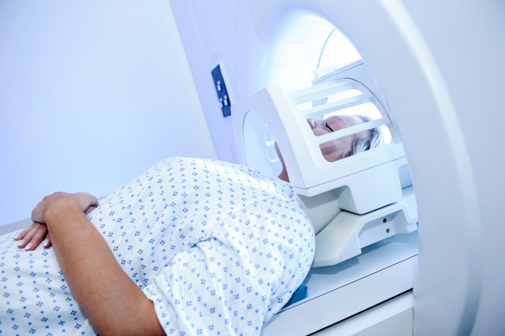
First things first. We’ve all heard of MRI scans and some of you already even had one or two in the past, but what is an MRI scan exactly? MRI stands for magnetic resonance imaging. This type of scan uses a large magnet, radio waves and a computer to create detailed, cross-sectional images of internal organs, tissues and structures within the body. The MRI scanner itself resembles a large tube with a table in the middle – for the patient to slide in.
Ever since the MRI was invented, it has been used continuously in the medical world. Experts are still working on refining the techniques of MRI to make sure that it can be of an even better assistance in medical procedures and research. The invention and the ongoing development of MRI scans are – without a doubt – revolutionary.
Nowadays, the use of MRI’s has become a very common procedure around the world. Also in the world of fillers! Because we have found a way to implement it into the flow of ARtery 3D. As a result, we have made it possible for doctors to visualize the arterial network of each individual patient through Augmented Reality. All it requires is a one-time, risk-free and harmless MRI scan.
What is it used for?

MRI scans are used for all sorts of things in the medical world. These are some examples in which MRI’s can really help by giving doctors, scientist and researchers the possibility to examine the inside of the human body in high detail:
► Tumors, cysts, anomalies of the brain and spinal cord or other anomalies in various parts of the body
► Heart problems (certain types)
► Breast cancer screening for women
► Uterine anomalies in women undergoing evaluation for infertility
► Liver diseases and diseases of other abdominal organs
► Back and knee injuries or abnormalities of other joints
► Evaluation of pelvic pain (women)
► …
Of course, this list of examples is far from complete. MRI technology is expanding in scope and use every single day and will continue to do so.
But there is one other example that we can definitely add to the list: filler treatments with ARtery 3D.
The flow: from MRI to ARtery 3D
Prescription
It’s very easy to print out a prescription in our ARtery 3D app to go get an MRI scan. All you need is the patient’s name and the date of birth. Here’s how it can go:
► The doctor performs some filler injections, but not all. Then he or she asks the patient to go get an MRI scan in order to perform the other ‘more risky’ injections in a safer way with ARtery 3D.
► The doctor performs the entire procedure and asks the patient to go get an MRI scan, so that he or she can use ARtery 3D to perform the filler injections with ARtery 3D upon the next visit.
This last situation can also be a perfect solution, if – you as a doctor – are getting the impression that the patient is dreading the scan.
Preparation
We start off with a 15-minute infrared lamp preparation without jewelry or make-up where the patient has to make a number of facial expressions. This is to enhance the blood flow.
At the MRI firm, the doctor may ask the patient to change into a gown and any metal jewelry or accessories should be removed. No worries though, if patients have metalwork – like dental implants – for example, only a little bit of shadow will be shown on the scan. So this won’t affect the use of ARtery 3D in any way. The MRI scans needed for our app are nothing like the conventional scans: they only take up to 12 minutes tops and no contrast agent is needed. The procedure is also non-invasive and painless.
During the scan
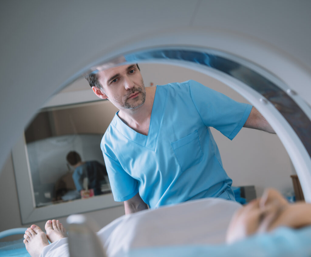
The doctor and the staff ensure that the patient is as comfortable as possible during the scan. Earplugs or headphones are provided to block out the noise from the MRI machine and the patient has to lay perfectly still. If necessary, they can communicate with the MRI technician via an intercom at all times.
After the scan
After the scan is done, the MRI center will upload the images in our app. Only in extremely rare cases, patients will experience side effects from an MRI scan, so no need to worry about that. This was it: the patient has undergone a one-time harmless scan and from now on any filler treatment in the future can be performed with ARtery 3D.
The benefits for your center
What’s in it for the MRI firms and radiology centers? Augmented Reality is a booming business in the medical environment, so it’s the perfect opportunity for your center to jump on board! But there are more benefits:
► Significant increase in revenue with a minimal effort on your side
► Continuous stream of patients being sent to your team to get the type of scan – required for ARtery 3D
► One-time implementation needed (and done in less than one hour!)
► Free training session provided with all the info you need
Our radiologists have collaborated with the 3 biggest MRI firms: GE, Philips and Siemens to develop this new 3D Time of Flight MOTSA sequence. It’s completely free and developed by Augmented Anatomy and it’s available on +80% of all 3T MRI’s worldwide.
